Home

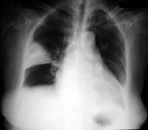
valley computer mosaic pulmonary embolism cxr wedge shaped dinosaur elevation Flavor

Hampton hump in acute pulmonary embolism | Cleveland Clinic Journal of  Medicine
Hampton hump in acute pulmonary embolism | Cleveland Clinic Journal of Medicine

Hampton hump in acute pulmonary embolism | Cleveland Clinic Journal of  Medicine
Hampton hump in acute pulmonary embolism | Cleveland Clinic Journal of Medicine

Initial and the follow-up chest radiographs of pulmonary infarction.... |  Download Scientific Diagram
Initial and the follow-up chest radiographs of pulmonary infarction.... | Download Scientific Diagram

Dark lung fields
Dark lung fields

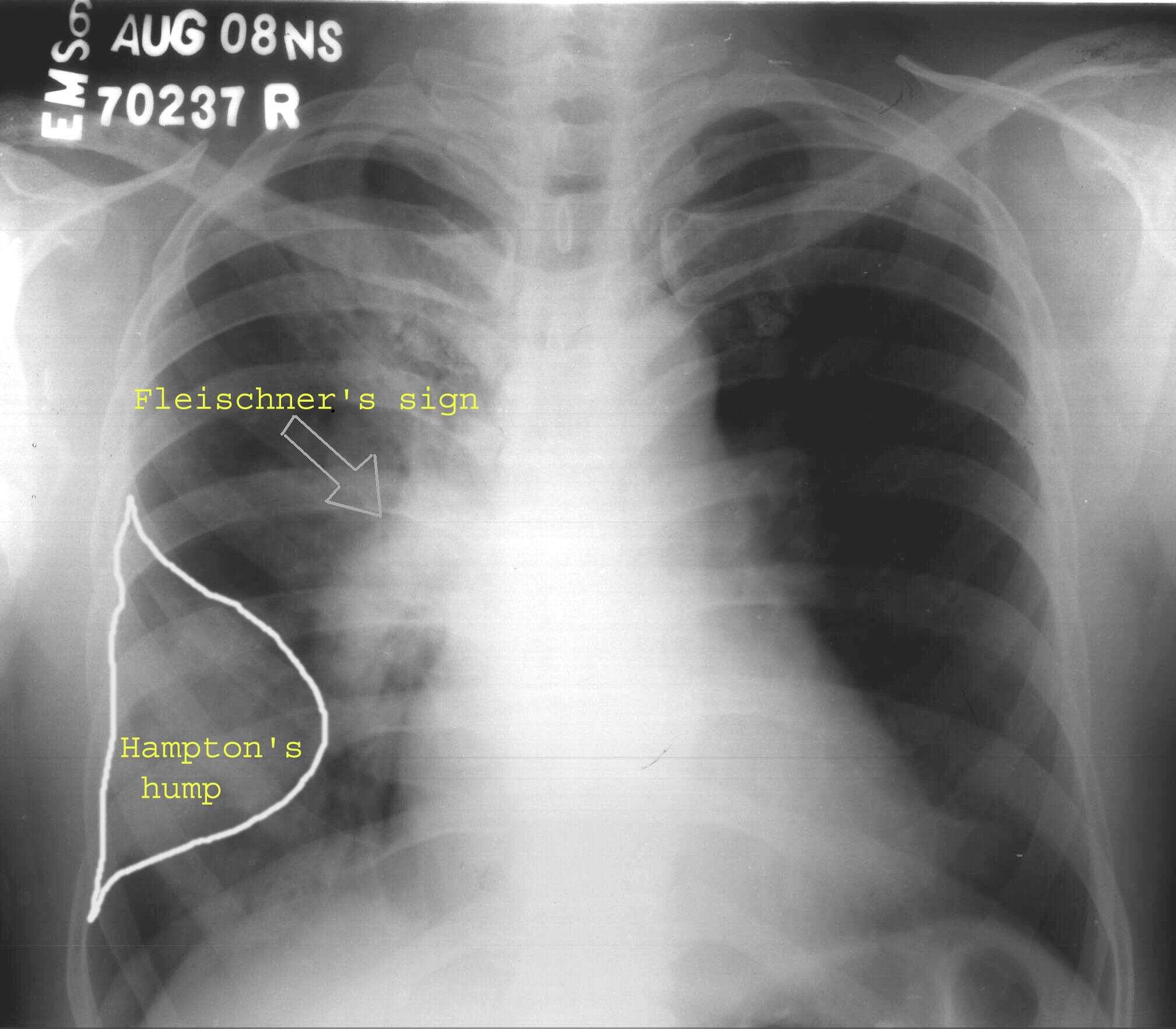
Hampton hump and Palla sign: Radiographic signs of pulmonary embolism |  Eurorad
Hampton hump and Palla sign: Radiographic signs of pulmonary embolism | Eurorad

Hampton hump | Radiology Reference Article | Radiopaedia.org

EPOS™
EPOS™

Peripheral wedge-shaped radiographic lung opacity in a young patient. -  Abstract - Europe PMC
Peripheral wedge-shaped radiographic lung opacity in a young patient. - Abstract - Europe PMC

Hampton's Hump | Imaging

Bilateral pulmonary emboli with Hampton hump sign | Radiology Case |  Radiopaedia.org
Bilateral pulmonary emboli with Hampton hump sign | Radiology Case | Radiopaedia.org

Pulmonary Embolism Diagnosis Guide | PPT

Pulmonary infarction | Radiology Reference Article | Radiopaedia.org

Hampton's Hump - pulmonary embolism - Radiology at St. Vincent's University  Hospital
Hampton's Hump - pulmonary embolism - Radiology at St. Vincent's University Hospital

Hampton hump | Radiology Reference Article | Radiopaedia.org

pulmonary infarction | pacs

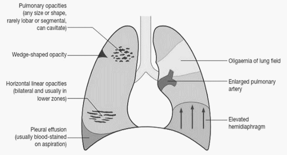
damsdelhi - BASICS - CHEST X-RAY IN PULMONARY EMBOLISM #rahulrajeev  #medicine A normal or near-normal Chest X-ray often occurs in pulmonary  embolism. Well established abnormalties include: • Enlarged pulmonary  artery-Fleishner sign •
damsdelhi - BASICS - CHEST X-RAY IN PULMONARY EMBOLISM #rahulrajeev #medicine A normal or near-normal Chest X-ray often occurs in pulmonary embolism. Well established abnormalties include: • Enlarged pulmonary artery-Fleishner sign •
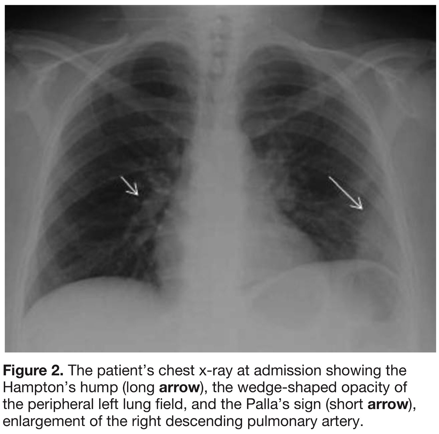
Edgar V. Lerma 🇵🇭 on X: "Pulmonary Embolism (PE) on CXR signs: ✓ Hampton  hump - Pleural-based wedge-shaped defect from infarction just above the  diaphragm ✓ Palla Sign - Large Right descending
Edgar V. Lerma 🇵🇭 on X: "Pulmonary Embolism (PE) on CXR signs: ✓ Hampton hump - Pleural-based wedge-shaped defect from infarction just above the diaphragm ✓ Palla Sign - Large Right descending

Southwest Journal of Pulmonary, Critical Care and Sleep - Imaging - Medical  Image of the Month: Hampton Hump and Palla Sign
Southwest Journal of Pulmonary, Critical Care and Sleep - Imaging - Medical Image of the Month: Hampton Hump and Palla Sign

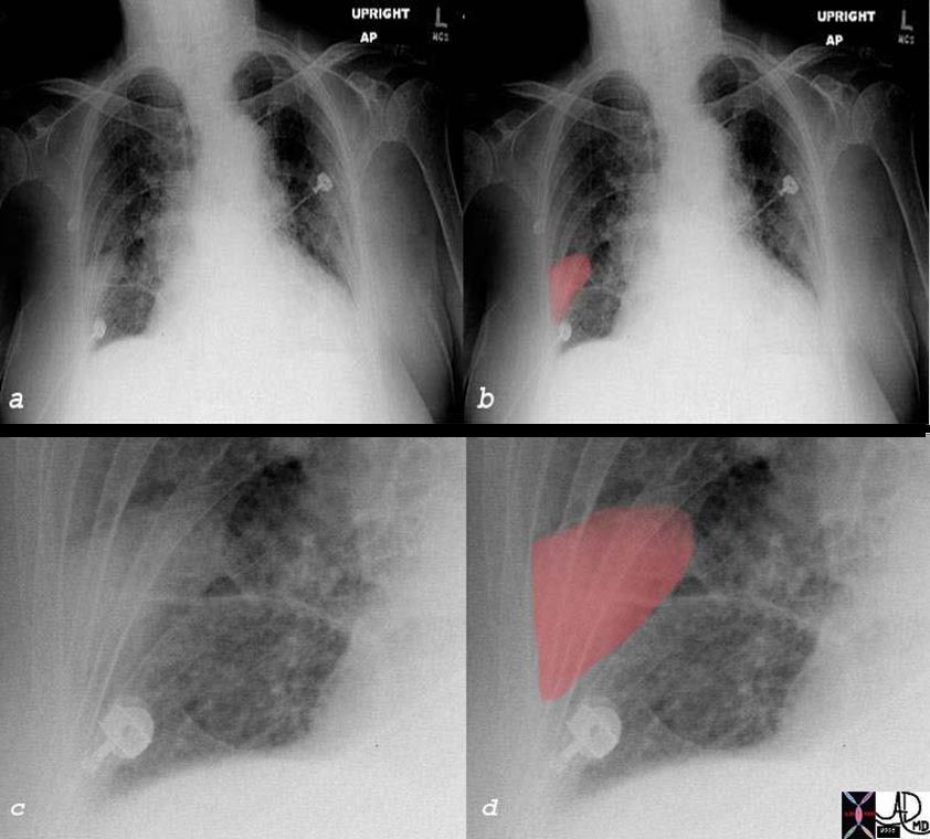
Figure, Wedge shape pulmonary infarction seen on AP chest x-ray. Image  courtesy of S. Bhimji MD] - StatPearls - NCBI Bookshelf
Figure, Wedge shape pulmonary infarction seen on AP chest x-ray. Image courtesy of S. Bhimji MD] - StatPearls - NCBI Bookshelf

Pin on Misc ...
Pin on Misc ...

Hampton hump | Radiology Reference Article | Radiopaedia.org

Follow up chest radiograph (day 5 of admission) showing wedge shaped... |  Download Scientific Diagram
Follow up chest radiograph (day 5 of admission) showing wedge shaped... | Download Scientific Diagram

Pulmonary Embolism : Chest X-ray Signs | Epomedicine

Radioloksabha - Articles

Chest radiograph of the patient in the case report revealing a... |  Download Scientific Diagram
Chest radiograph of the patient in the case report revealing a... | Download Scientific Diagram

Case 5: HRCT showing bilateral, wedge-shaped peripheral opacities (see... |  Download Scientific Diagram
Case 5: HRCT showing bilateral, wedge-shaped peripheral opacities (see... | Download Scientific Diagram